

Overview
The Histopathology Core Facility is one of six Core Facilities that are central to the Unit’s research studying how cells, tissues, and organisms respond to different types of injury, triggered by environmental agents and endogenous molecules. We provide histopathology technical services, advice, and training to scientists at all levels.
Services
We offer a high throughput formalin fixed paraffin embedded (FFPE) histology service for tissue samples from human, mouse, and other model organisms. Using our well-equipped and highly automated Histopathology laboratory, our staff can provide a comprehensive range of histopathological techniques including:

- Tissue fixation
- Grossing (initial examination, description and selection of optimal region)
- Sample preparation
- Tissue processing into FFPE blocks
- Microtomy (tissue sectioning)
- Tinctorial slide staining (selective or special stains).
We also undertake a wide range of highly quantitative and complex in situ assays, including fully automated multiplex immunohistochemistry and co-in situ hybridisation.
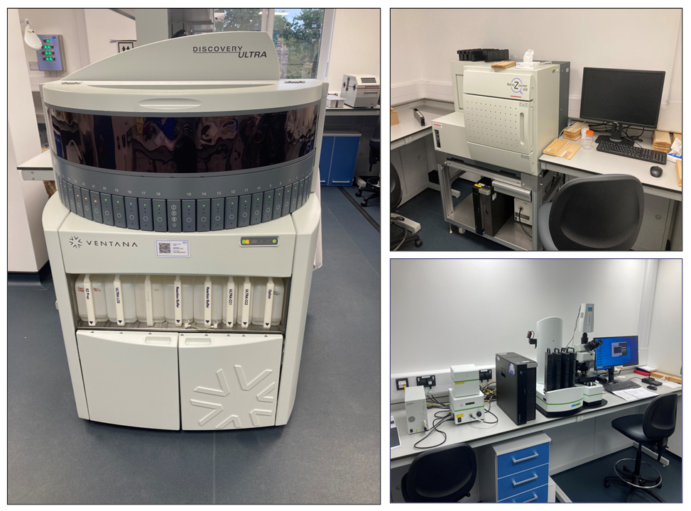
Key instrumentation
- Leica Pearl Tissue Processor
- A range of microtomes and vibratomes
- Automated Haematoxylin and Eosin staining and coverslipping.
- Roche Discovery Ultra Immunostainer for chromogenic and multiplex immunofluorescence
- Hamamatsu Nanozoomer Slide Scanner for brightfield whole-slide imaging
- Akoya Vectra3 digital imaging system for immunofluorescence whole slide imaging and spectral unmixing
Together these resources can be used to explore protein and RNA expression in a wide range of tissue samples. We also support and facilitate image analysis service across multiple platforms, including Akoya Vectra InForm and Spatial Biology using Visiopharm.
Facility staff
|
Histopathology Manager: Mark Southwood |
Histology Research Officer: Emma Gould |
Academic Lead: Marion MacFarlane |