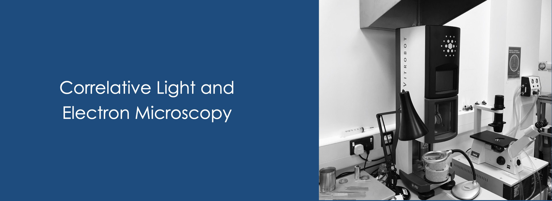
Correlation Light and transmission or scanning Electron Microscopy for low expression proteins.
Coverslip options:
- Photoetch gridded coverslip for chemical fixation (ibidi, matsunami)
- Gridded glass-bottom dish by photo-etching for chemical fixation (ibidi, matsunami)
- Gridded sapphire coverslip by Quorum K950X Turbo Evaporator with Leica CLEM coating mount
Workflow for RT-CLEM (LM before the plastic embed):
- Culture tissue cells on the gridded coverslip, (in which the fluorescent proteins are expressed)
- Observe living cells by confocal fluorescence microscopy with gridded patterns
- Chemical fixation with half-strength Karnovsky’s solution (2% paraformaldehyde - 2.5% glutaraldehyde, EM-grade) in sodium cacodylate buffer including 2mM CaCl2 or NaHCa buffer
- Rapid freeze tissues or cultured cells by Leica EM-HPM100 Sapphire disc system or Variant CryoPress (as required)
- Freeze substitution by Leica EM AFS2 with FSP (as required)
- Infiltration and resin embedding in epoxy resin by Leica EM-TP
- Polymerization at 70 °C for a few days by (vacuum) oven
- Remove the coverslip to leave the gridded pattern
- Trim the plastic block to identify the correlated same field
- Semi-thin section and stain with 1% toluidine blue solution
- Ultrathin section by Leica EM UC7 or ARTOS-3D
- Grid stain with uranyl acetate (or alternative) and lead citrate
- Correlation imaging by TEM or SEM
Workflow for In-resin CLEM (LM after the plastic embed):
- General procedures for high-pressure freezing and freeze substitution
- Confocal fluorescence microscopy fluorescent molecule-tagged protein expressed cells in Lowicryl HM20 ultra-sections
- Grid stain with uranyl acetate (or alternative) and lead citrate
- Correlation imaging by TEM or SEM
Workflow for Cryo-CLEM:
- Confocal fluorescence microscopy living GFP-tagged protein expressed cells on gold EM grids
- Cryo-Confocal fluorescence microscopy (as required) [under construction]
- Plunge freeze in liquid ethane or ethane/propane mixture by the TFS Vitrobot
- Cryo-EM imaging


