
Cryo-Electron Microscopy of Vitreous Sections (CEMOVIS) for nanoscale molecular structures.
Workflow:
- Vitrification tissue cells by Leica EM HPM100, TFS Vitrobot, or plunge freezing in super cooled slush nitrogen
- Set onto the specimen pins for cryo-ultramicrotomy
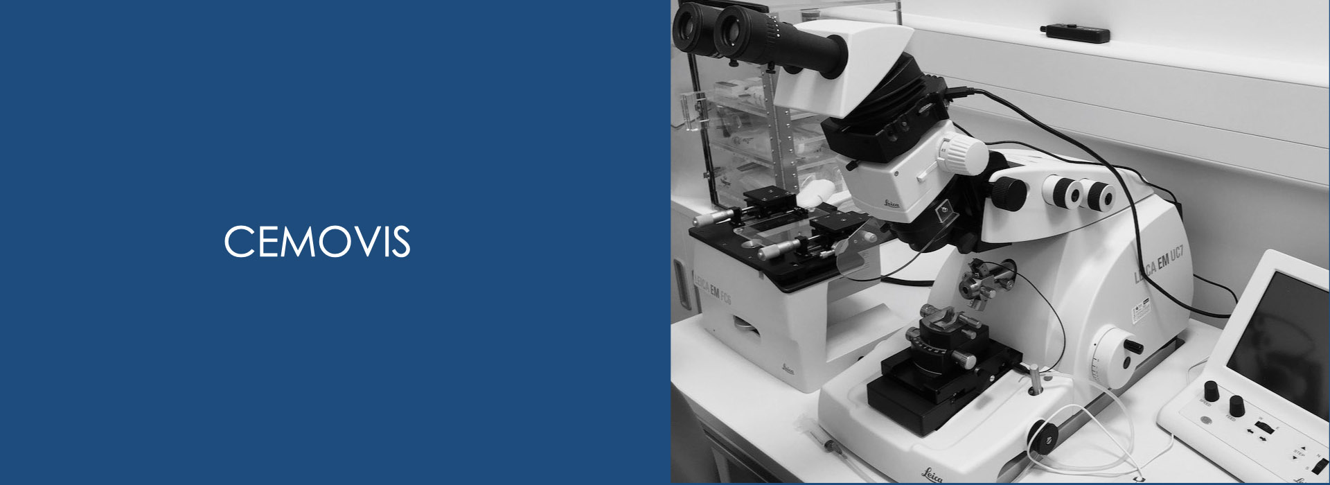
- Cryo-trim the frozen block with Diatome cryo-trim diamond knife (35°) by Leica EM UC7-FC7 at liquid nitrogen temperature
- Cryo-ultra-section with Diatome cryo-immune diamond knife (35°) and making ribbons by Leica EM Crion
- Collection the frozen ribbon on the grid by Leica EM double-manipulator
- Transfer the cryo-grids to the cryo-storage box
- Set into the cryo-transfer holder
- General alignments for cryo-EM
- Low-dose imaging by cryo-EM and tomography
- Analysis for nanoscale molecular structures