
Overview
The MRC Toxicology Unit has a long track record of using X-ray crystallography, nuclear magnetic resonance (NMR) spectroscopy and electron microscopy (EM) to obtain structures that provide novel mechanistic understanding that links exposure to adverse outcomes.
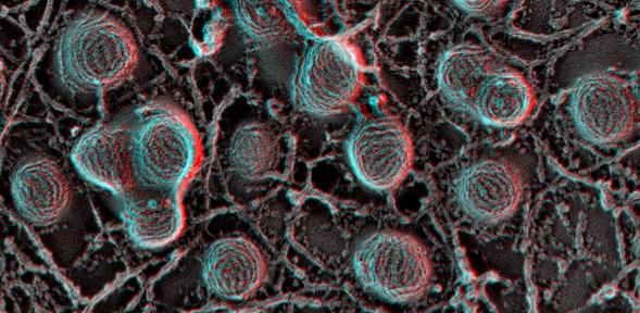
The Electron Microscopy & Ultrastructural Pathology (EMUP) Core Facility provides three-dimensional and large-volume imaging analysis at EM resolution for the MRC Toxicology Unit core programmes and the external collaborative projects. This is achieved using an integrated EM for both cryogenic (cryo) and room temperature (RT) and a variety of different kinds of sample preparation devices for cryo-vitrification (high-pressure, mirror and plunge freezing), fine carbon coating, glow-discharging, plasma cleaning, deep etching, gold sputtering and low-angle rotational shadowing with fine platinum.
Our nano-toxicology approach at the EMUP facility enables the study of a whole range of tissues and cultured cells from mouse lung, drosophila gut, microbiome, mesothelioma, lung carcinoma, and cardiomyocytes to filamentous protein-complexes. The designed protocol for an integrated EMUP can be specialized based on the type of your biological specimen.
Services
The EMUP Facility for Cryo- and RT-challenges offers the whole range of procedures from sample preparation to image acquisition. This encompasses a complete service, including an initial meeting, which is essential for identification of the most effective EM solution, experimental designs, and time schedules.
Correlative changes between structure and function can be examined in nanoscale to enable an in-depth visualisation of areas adversely affected by toxins or drugs. This is done by using:
- 3D-Transmission Electron Microscopy (TEM) - using electron tomography and single particle analysis
- Volume Scanning electron microscopy (SEM) – involving serial block face imaging
- Cryo-techniques.
Through a technical development for challenging biological specimens, the MRC Toxicology Unit EMUP Facility has well designed advanced techniques for EMUP as well as conventional TEM/SEM.
Designed techniques conducted at the EMUP Facility
Sample preparation
- Chemical fixation, osmication, block staining, infiltration, dehydration, embedding, fine trimming and ultra sectioning
- Vitrification (rapid freezing) by high-pressure or plunge freezing
Conventional 2D snapshot
- Conventional ultra-cut TEM for chemical fixation and epoxy embedding
- Conventional SEM for chemical fix, HMDS and gold sputtering
3D and large-volume imaging
- Single particle analysis and negative staining TEM
- TEM/HAADF-STEM tomography
- Serial-block-face SEM with 3View system
- Cryo-Electron Microscopy of Vitreous Sections (CEMOVIS)
Advanced preparation for challenging materials
- High-pressure freezing and freeze-substitution (FS)
- Immune-gold labelling for FS Lowicryl HM20/K4M
- Classic types of correlation light and electron microscopy with FS
- Rapid-freeze, deep-etch and freeze fracture EM
Key equipment
RT-EM Imaging Suite (Gleeson building)
- JEOL JEM1400HC TEM/TVIPS TemCam-XF416
- FEI Quanta 250FEG SEM
- Gatan 3View System/OnPoint BSE Detector
- Quorum GloQube Glow Discharge System, K950X Turbo Evaporator
- Leica EM Artos3D, TRIM2, TP, KMR3, ACE200
Cryo-EM Suite (Pharmacology building)
- FEI TalosF200C/Ceta16M/Falcon2/HAADF-STEM detector/Vitrobot
- Gatan Cryo-Transfer Holder 626/914, Turbo Pumping Station
- Fischione Advanced Tomography Holder (RT) 2020
- Leica EM HPM100, AFS2/FSP, EM FC7/UC7/Manipulator/CRION
- JEOL JFDII
Imaging Tech Office (Gleeson building)
- Workstation for Amira3D (Precision 5820 Tower XCTO Intel Xeon Processor W-2275 14C 3.3GHz, 256GB RAM, Nvidia RTX A6000 GPU 48GB, 12TB HD 1TB SSD)
- Workstation for EMAN2/IMOD4(5), Inspect3D/Argos (5820T, RedHat Linux, Nvidia RTX A6000 GPU 48GB, 12TB HD 1TB SSD)
- Cryo-EM Server for Relion4(5) (PowerEdge T550 Server 2x Intel Xeon Gold 6326 2.9GHz 16C/32T, 256GB RAM, Nvidia Ampere A40 GPU 48GB, 7.68TB SSD), operated by the Bioinformatics Core facility
Facility staff
|
Facility Head and Academic Lead |
Selected Publications
Hardy RE, Chung I, Yu Y, Loh SHY, Morone N, Soleilhavoup C, Travaglio M, Serreli R, Panman L, Cain K, Hirst J, Martins LM, MacFarlane M, Pryde KR. (2023) The antipsychotic medications aripiprazole, brexpiprazole and cariprazine are off-target respiratory chain complex I inhibitors. Biol Direct. 18(1):43.
Grosso S, Marini A, Gyuraszova K, Voorde JV, Sfakianos A, Garland GD, Tenor AR, Mordue R, Chernova T, Morone N, Sereno M, Smith CP, Officer L, Farahmand P, Rooney C, Sumpton D, Das M, Teodósio A, Ficken C, Martin MG, Spriggs RV, Sun XM, Bushell M, Sansom OJ, Murphy D, MacFarlane M, Le Quesne JPC, Willis AE. (2021) The pathogenesis of mesothelioma is driven by a dysregulated translatome. Nat Commun. 12(1):4920.
Fox JL, Hughes MA, Meng X, Sarnowska NA, Powley IR, Jukes-Jones R, Dinsdale D, Ragan TJ, Fairall L, Schwabe JWR, Morone N, Cain K, MacFarlane M. (2021) Cryo-EM structural analysis of FADD:Caspase-8 complexes defines the catalytic dimer architecture for co-ordinated control of cell fate. Nat Commun. 12(1):819.
Buckley N, Panatta E, Morone N, Noguchi M, Scorrano L, Knight RA, Amelio I, Melino G. (2020) P73 C-terminus is dispensable for multiciliogenesis. Cell Cycle. 19(14):1833-1845.
Amelio I, Panatta E, Niklison-Chirou MV, Steinert JR, Agostini M, Morone N, Knight RA, Melino G. (2020) The C terminus of p73 is essential for hippocampal development. Proc Natl Acad Sci USA. 117(27):15694-15701.
Song Y, Dagil L, Fairall L, Robertson N, Wu M, Ragan TJ, Savva CG, Saleh A, Morone N, Kunze MBA, Jamieson AG, Cole PA, Hansen DF, Schwabe JWR. (2020) Mechanism of crosstalk between the LSD1 demethylase and HDAC1 deacetylase in the CoREST complex. Cell Rep. 30(8):2699-2711.e8.
Morone N, Usukura E, Narita A, Usukura J. (2020) Improved unroofing protocols for cryo-electron microscopy, atomic force microscopy and freeze-etching electron microscopy and the associated mechanisms. Microscopy (Oxford). 69(6):350-359. doi: 10.1093/jmicro/dfaa028.
Peretti D, Bastide A, Radford H, Verity N, Molloy C, Martin MG, Moreno JA, Steinert JR, Smith T, Dinsdale D, Willis AE, Mallucci GR. (2015) RBM3 mediates structural plasticity and protective effects of cooling in neurodegeneration. Nature. 12;518(7538):236-9. doi: 10.1038/nature14142.
Tufi R, Gandhi S, de Castro IP, Lehmann S, Angelova PR, Dinsdale D, Deas E, Plun-Favreau H, Nicotera P, Abramov AY, Willis AE, Mallucci GR, Loh SH, Martins LM. (2014) Enhancing nucleotide metabolism protects against mitochondrial dysfunction and neurodegeneration in a PINK1 model of Parkinson's disease. Nat Cell Biol. 16(2):157-66. doi: 10.1038/ncb2901.
Sinha B, Köster D, Ruez R, Gonnord P, Bastiani M, Abankwa D, Stan RV, Butler-Browne G, Vedie B, Johannes L, Morone N, Parton RG, Raposo G, Sens P, Lamaze C, Nassoy P. (2011) Cells respond to mechanical stress by rapid disassembly of caveolae. Cell. 144(3):402-13.