A tissue microarray is a single formalin fixed paraffin embedded (FFPE) block containing cores from multiple FFPE samples. The use of Tissue Micro Arrays (TMAs) enables the simultaneous application of histological and in-situ assays such as immunohistochemistry (IHC) and in-situ hybridisation (ISH) to multiple tissue samples simultaneously, a single TMA can contain over 300 cores. A TMA can be a valuable resource when working with archival specimens as a small representative sample is taken, preserving the larger FFPE block for future use. They can also be used to investigate a panel of tissue, answering broader questions whilst using fewer reagents.
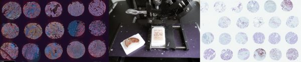
Once FFPE cases or specimens have been identified, slides are stained with haematoxylin and eosin (H&E) and a full slide image is captured with the Hamamatsu Nanozoomer. Slides are then reviewed to identify and annotate target areas of tissue or tumour for TMA construction. A TMA map is then designed to show the location and identification number of each core.
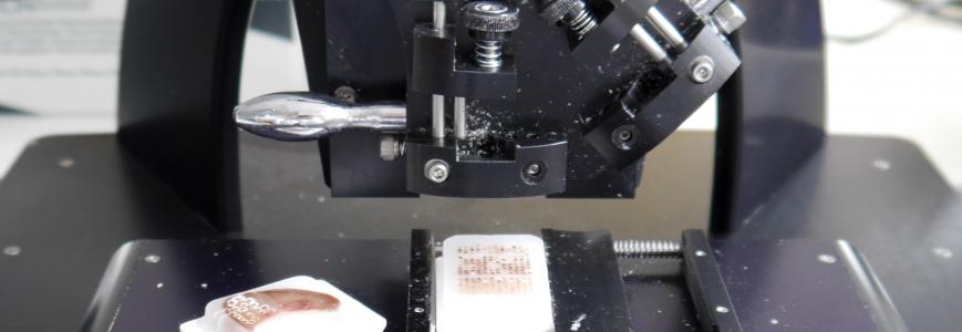
Using our semi-automated TMArrayer, a ”blank” wax donor block is prepared by creating appropriately sized (1mm-2mm) holes for the TMA cores in the pattern of the TMA map. Cores are then taken from the identified areas and placed into the corresponding receptacle hole. Once complete the block is placed in an incubator to set then can be sectioned as a normel FFPE block. Sections can be used for any tissue based assay and can produce vast quantities of quantifiable data.