Our Science-Art prizes are awarded to the best creative entry that tells the story of the research that we do at the Unit. Entries can be a microscopy image, a photograph, a video or any other creative endeavour. Two prizes are awarded annually. The judges choice winner is selected by a judging panel made up of a scientist, science communicator and an artist, and our audience winner is selected by a popular vote including all Unit members.
2023
| Untangling the rose with chemical thorns: Untangling the rose with chemical thorns” represents the duality and contradiction from how chemicals which have been central to modern life development can also be toxic to us. In my job I am trying to untangle how we are getting caught on the “thorns”, specifically through finding the mechanisms behind the effect of toxic xenobiotics on the human gut microbiome. This would allow us to mitigate the effect of toxic xenobiotics in our health and develop safer chemicals. The image also invites us to rethink the rose of modern production systems. | |
|
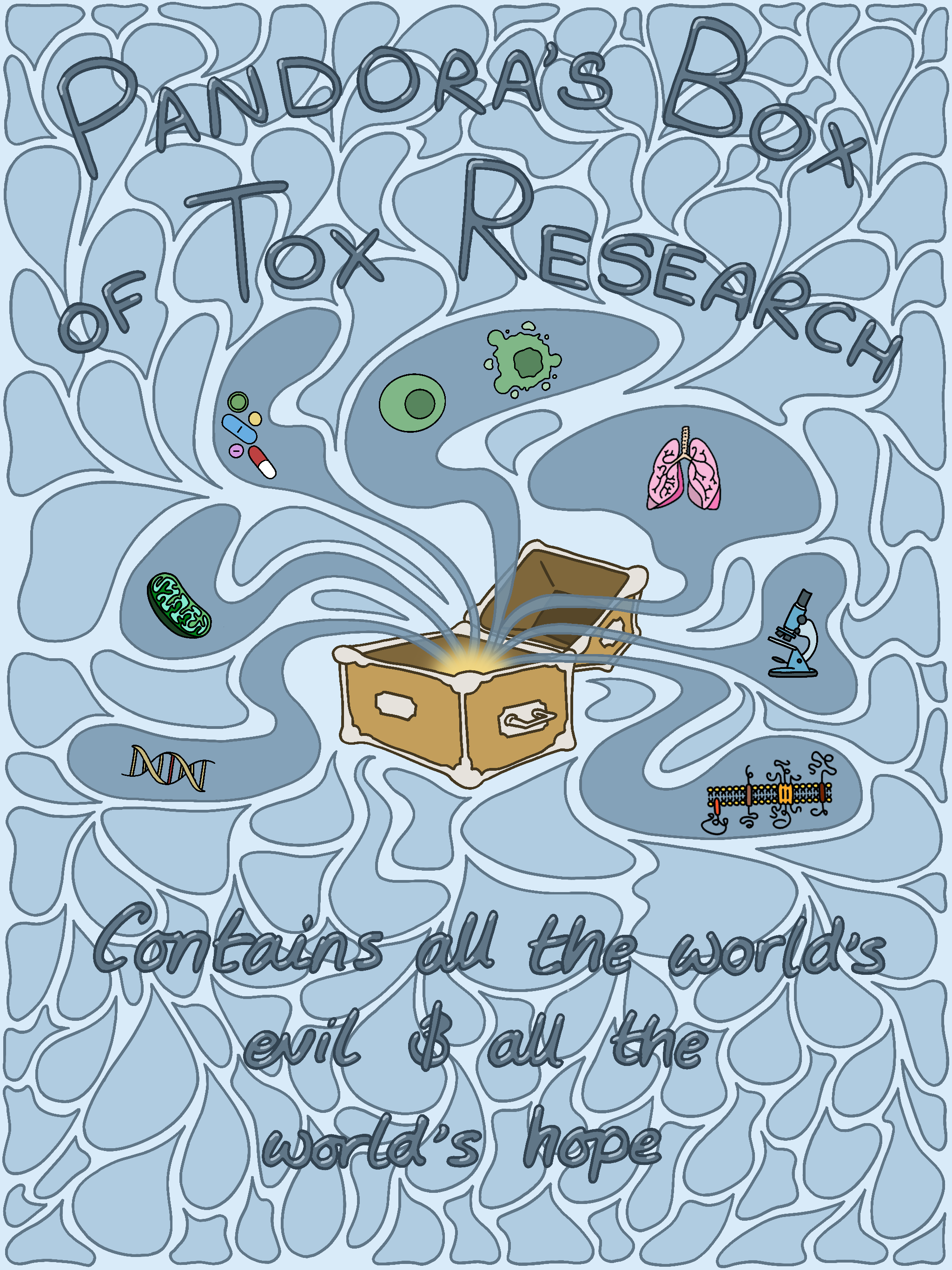
Pandora's Box of Tox Research: I see toxicology research as a Pandora's Box which is traditionally known to "contain all the world's evils and all the world's hope." At the MRC Toxicology Unit, we study things like air pollution and brake/tyre wear, which can feel like the world's evils because of their multifaceted impacts on health. However, toxicology research can provide hope and solutions in the form of therapeutics to counter the negative impacts of these toxicants. |
|
|
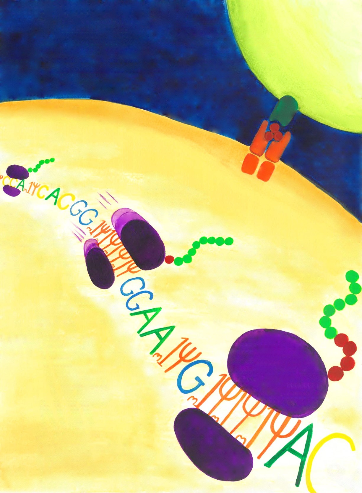
Ribosome frameshifting on modified mRNAs: This image illustrates the findings of work by Tom Mulroney in the Willis lab. Tom found that (N)1-methylpseudouridine incorporation into mRNA, a modification used in mRNA vaccines, results in +1 ribosomal frameshifting sites where the ribosome can slip and begin translating a frameshifted protein. |
|
|
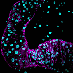
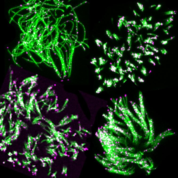
ACE at the tips: As part of our research into the mechanisms of toxicity and disease in the cells lining our airways we studied the distribution of the ACE2 receptor in the multi-ciliated cells that form part of this lining. This receptor, shown in magenta, is part of the entry route for the virus responsible for Covid-19 disease and is seen at the tips of the cilia, shown in green. The image is an assembly of 4 different cells, illustrating the variety of cilia orientations and lengths. |
|
|
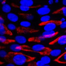
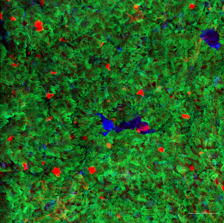
Cilia carpet: We grow human airway cells on a porous membrane, exposing the cells to air on one side and to a differentiation medium on the other side. After about one month a large number of cells become studded with cilia, hairlike projections shown here in green. Studded among the ciliated cells are secretory cells, shown in red, and precursor cells that can differentiate into either cell type, shown in blue. Together these cells are responsible for clearing pathogens and toxicants from our airways. |
|
|
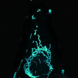
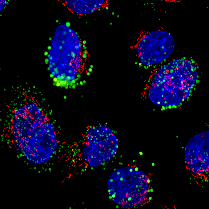
Lipid droplets: While they are sometimes cast in a bad light, lipids are essential for cell health. In these human airway stem cells, lipids are stored in numerous lipid droplets, shown in green. When grown at an air-liquid interface these cells can differentiate into multi-ciliated cells which together with secretory cells are responsible for clearing our airway of mucous. In this process of differentiation the lipids stored in the stem cell droplets are used to make the extra cell membrane required to create the cilia. Also shown are the cell nuclei in blue and the Golgi apparatus in red. |
|
|
My lab in drawings: What makes a lab group a lab group? What are some of the common ideas, tools, and specimens we work with? In this drawing, I aimed at summarising what the Patil Group does; including both laboratory and computational touches. Our lab does wide ranging work, from laboratory experiments on yeast evolution, to gut bacterial interactions with xenobiotics and drugs, to metagenomics and mathematical modelling. In this drawing, I intended to merge all of these facets so that the diversity of our lab's work comes to light. |

2022
| Slice of Life: Amrita's ink drawing is made using thousands of little dots (pointillism) depicting a slice through a cell. The image focusses on different aspects of mitochondria, it's transport and interaction with other organelles, fission-fusion and undergoing destruction (mitophagy). Mitochondria is one of the major organelles that are affected by toxins and sitting at the centre of cellular biochemistry, plays a very important role in life, death and recovery of cells. (Winner) | |
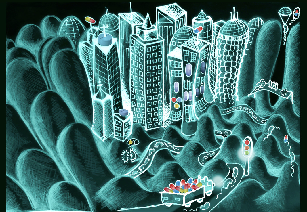
| The Big Bug Hub: Naomi drew a cartoon called 'Bug Hub': the inside of our guts, in particular the epithelial cells that are like skyscrapers in big cities for our gut microbiota. The background was drawn by hand and the cartoon drawn digitally over it.
With this cartoon, I am trying to make the metaphor that the gut may seem like a big, chaotic city at first, especially when exposed to 'external' things like drugs (i.e. unexpected, incoming traffic). Like in a big city, there is lots of stuff happening at various scales. However, if we learn when and where to zoom in, and equally, when to take a step back, patterns of organisation and order may appear. (Winner) |
|
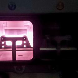
| Trooping the Colour: In-air glow discharge is a technique for cleaning and modifying surfaces in electron microscopy sample preparation, leaving the TEM grids’ surface hydrophilic and negatively charged to anchor the sample and support staining for good imaging contrast. Air contains trace amounts of noble gases; when an electrical discharge passes through a noble gas at low pressure, the gas will glow due to collisions between molecules in the gas and electrons of the current that leaves the air partially ionized. When this phenomenon occurs naturally in stormy weather, we call it St. Elmo’s Fire; when it occurs inside a glass tube, we call it a neon sign. At the EM facility we house the Quorum GloQube to help in the task. In scientific remembrance of late Queen Elizabeth II, an old Lady who enjoyed an extraordinary life! | |
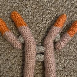
| Crocheted Antibody: In our lab, we investigate the impact of immune checkpoint blockade therapies on the immune response to vaccination. This crocheted antibody (created using the Antibuddy pattern from @adele_valeria) shows the basic structure of the specific antibodies that can be formed in response to vaccination. The production of these antibodies can be impaired in people on immune checkpoint blockade due to the expansion of a subset of B cells known as Age-Associated B cells. | |
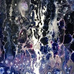
| A tricky UMAP: I’m currently collaborating on a project that is trying to disentangle progressive cell differentiation in early embryogenesis in mice. I wanted to create something that reflected the progression of cell identity whilst also showing how ambiguous the borders between cell types can be. Mixing layers of acrylic paints of different densities with silicon oil creates these shapes as the oil and water components separate and the paints slide and bubble past each other. This process continues as the moisture evaporates and the painting continues to change until the final stage when it is fully dry. | |
| River of Sorrow: The digestive tract encounters toxins on a daily basis. We use Drosophila as a model system to look at the damaging effects of different drugs on the intestinal cells. This picture depicts the gut cells with active caspases which are cell death proteins (in magenta) and nuclei (in cyan) upon ingestion of toxins. | |
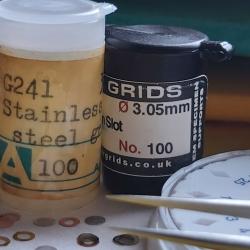
| Geometry of the Naked Grid: A TEM grid is typically a flat disc with a mesh or other shaped holes used to support ultrathin sections of specimens for electron microscopy imaging, an important tool in biomedical research to investigate the detailed structure of tissues, cells, organelles and macromolecular complexes upon toxic insult and disease. Grids are available in a wide variety of hole patterns (squares, hexagons, bars, slots) and materials (gold, copper, nickel, titanium, stainless steel, aluminium). Specimens might require an additional support on top of the grid to prevent them from falling through the holes: carbon coating and glow discharge provide support for imaging without interfering with the flow of the electron beam. The digestive tract encounters toxins on a daily basis. We use Drosophila as a model system to look at the damaging effects of different drugs on the intestinal cells. This picture depicts the gut cells with active caspases which are cell death proteins (in magenta) and nuclei (in cyan) upon ingestion of toxins. | |
| Macrophages the Heroes: Long pathogenic fibres are retained in the cells during many years and eventually triggering the development of cancer. Macrophages fight against these fibres to eliminate them from our cells, however some fibres will be cleared, and some will escape from phagocytosis. These images from Perl Prussian Blue staining show the battle. | |
|
PhD: Day in the Life: The video depicts a relatively standard day in my life as a PhD student, detailing my toxicological research from bench to desk, concisely edited and synced to some music. |
|
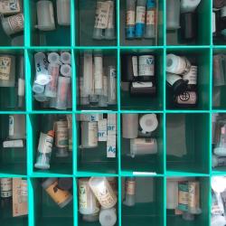
| I am not there (yet): The storage of TEM grids and resin embedded specimen blocks are an important aspect in sample processing in any electron microscopy laboratory. Archives are important because they provide evidence of past research activities and preserve significant information that might complement current research, providing a base knowledge upon which built on over time. A wide range of storage boxes for short term and archiving storage are commercially available. The image shows one the oldest grids storage box kept at the EM facility; the Tim’s box. For sensitive samples and grids which need protection against oxygen, humidity and contamination we have modern acrylic desiccator cabinets. | |
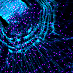
| Web of actin: The image shows part of a crop leading to the anterior midgut in Drosophila melanogaster, a common model system in toxicology research. The crop is similar to the mammalian stomach and undergoes contractions that help move food into the midgut for digestion. Toxins ingested can cause damage to gut cells but can also indirectly affect other organ systems including the brain. In this image, actin in muscles surrounding the crop have been stained using Phalloidin (cyan) and the cell nuclei using Hoechst (magenta). | |
| For your eyes only: The digestive tract contains acid secreting cells that help maintain an acidic environment in the gut. The picture depicts the acid secreting gut cells marked by the homeobox protein Cut (in red) and all cell nuclei labelled with hoechst (in blue). The expression of Cut around the cell nuclei gives it the appearance of eyes. | |
| Flaming Mitochondria: Mitochondria play essential roles in regulating cellular functions. Some drug treatments and molecular interventions have been reported to have off-target effects damaging mitochondria and causing severe side effects. Some of these drugs can cause mitochondria to "burn out". My image is a confocal image of mitochondria, which I added some more flames so that it looks cooler. |